Neurosurgical Focus · 2018

Manjila-Semaan Classification

A Standardized System for Jugular Bulb Positions
and Microsurgical Implications in Skull Base Surgery

Dr. Sunil Manjila, MD & Prof. Maroun Semaan, MD

Scroll to explore

About Dr. Sunil Manjila

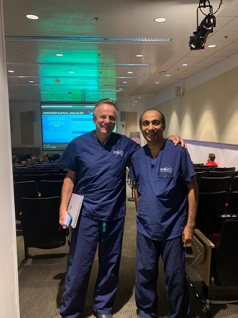
Dr. Sunil Manjila with Professor Robert Spetzler at Barrow Neurological Institute, Phoenix, Arizona, 2017
With Prof. Robert F. Spetzler · Barrow Neurological Institute (BNI), Phoenix, AZ - 2017

Dr. Sunil Manjila is a Michigan-based neurosurgeon whose career reflects a rare blend of rigorous training, innovative thinking, and clinical impact. His medical career began at Government Medical College, Thrissur, Keralam State of South India (Batch of 1989), inspired by a personal loss that shaped his calling to neurosurgery.

He completed his MCh (5-year program) with DNB in Neurosurgery at Christian Medical College (CMC), Vellore — one of Asia's most prestigious neurosurgical training centers — earning the Thiru Dr C V Chari Prize in Neurosciences. A short-term clinical fellowship in skull base and neurovascular surgery at Fujita Health University, Japan (2003). In 2004, he moved to the United States to pursue higher training. He subsequently undertook a full neurosurgical residency at Case Western Reserve University, Cleveland, with an enfolded fellowship.

In the US, a postdoctoral fellowship at Harvard Medical School in robotic cranial neurosurgery research led to the development of novel MR-compatible endoscopes. His pioneering work on robotics within the MRI bore has led to seminal scholastic publications. Encounters with legends Dr. Robert Spetzler and Dr. Neil Martin reinforced his commitment to systematic, reproducible neurosurgical frameworks.

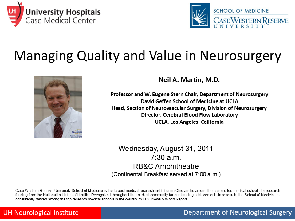
Grand Rounds Presentation by Dr. Neil A. Martin, MD at Case Western Reserve University, 2011
Presentation by Special Guest and Grand Rounds Speaker Dr. Neil A. Martin, MD · Case Western Reserve University, 2011

The Spetzler-Martin arteriovenous malformation (AVM) grading system, proposed in 1986, assigns points for various angiographic features of intracranial arteriovenous malformations to determine a score that predicts the morbidity/mortality risk associated with open surgery (https://thejns.org/view/journals/j-neurosurg/65/4/article-p476.xml). Outside the Glasgow Coma Scale (GCS) used in neurotrauma, Spetzler-Martin grading is the most popular grading system used in cranial neurosurgery.

The Classification System

A 5-tier system stratifying jugular bulb positions using high-resolution CT imaging, with sub-categories based on the presence or absence of dehiscence. Click a type to explore.

Legend — Anatomical Landmarks

  • IAC Internal Auditory Canal
  • PSCC Posterior Semicircular Canal
  • SCC Semicircular Canal
  • SPS Superior Petrosal Sinus
  • JB Jugular Bulb
  • ICA Internal Carotid Artery
  • I Incus
  • M Malleus
TABLE 1. Manjila and Semaan classification of jugular bulb location
Type Description
Type 1 No jugular bulb
Type 2 Below inferior margin of posterior SCC
Type 2a Without dehiscence into the middle ear
Type 2b With dehiscence into the middle ear
Type 3 Between inferior margin of posterior SCC & inferior margin of IAC
Type 3a Without dehiscence into the middle ear
Type 3b With dehiscence into the middle ear
Type 4 Above inferior margin of IAC
Type 4a Without dehiscence into the IAC
Type 4b With dehiscence into the IAC
Type 5 Combination of dehiscences

The 2018 Landmark Paper

Neurosurgical Focus 2018 · Volume 45, Issue 1 PMID: 29961385

"Jugular Bulb and Skull Base Pathologies: Proposal for a Novel Classification System for Jugular Bulb Positions and Microsurgical Implications"

Manjila S, Bazil T, Kay M, Udayasankar UK, Semaan M

This paper addressed a long-standing ambiguity in skull base surgery: the absence of a universally accepted definition for high-riding jugular bulb (HRJB). Different specialties — radiology, neurotology, and neurosurgery — used variable criteria, leading to miscommunication and surgical risk. The Manjila-Semaan system used high-resolution CT imaging to stratify jugular bulb positions into five major types with sub-categories based on dehiscence, directly aligning radiological anatomy with operative decision-making and enabling clear, reproducible interdisciplinary communication.

Video Overview

A complete walkthrough of the Manjila-Semaan Classification System — its rationale, anatomical basis, and surgical implications.

Key Publications Cited

Since its publication, the Manjila-Semaan Classification has been validated across multiple independent clinical and academic studies all over the world.

2023

Inter- & Intrarater Reliability Study

Journal of Neurological Surgery Part B: Skull Base

Demonstrated high inter- and intrarater reliability of the classification system across varying levels of radiological experience — a critical milestone for widespread clinical adoption in surgical planning.

Read Study →
2025

Vestibular Schwannoma Surgery Application

Neurosurgical Review · Springer

Applied the classification to preoperative CT evaluations in patients undergoing vestibular schwannoma excision, confirming that Type 4 jugular bulbs significantly increase the risk of venous injury during drilling — enabling proactive surgical planning.

Read Study →
2025

Validation of a Novel Jugular Bulb Classification in Otology & Lateral Skull Base Surgery: A Neuro-Otologist's Perspective

The Journal of Laryngology & Otology · 2025;139(10) · Das NK, Keshri A, Manogaran RS, et al.

A neuro-otology team at SGPGIMS, Lucknow independently validated the classification for otologic and lateral skull base surgery, confirming its reliability in preoperative surgical planning.

Read Study →
2026

Jugular Bulb Position Variability on Temporal Bone HRCT

J Coll Physicians Surg Pak (JCPSP) · 2026;36(1):20–24 · Uddin MMZ, Jamil A, Hashmi SQ, et al.

Applied the classification to HRCT temporal bone imaging in a Pakistani population, characterising jugular bulb type variability across age groups.

Read Study →
2024

Jugular Bulb Variants on HRCT — Imaging & Clinical Correlation

Medicina (Kaunas) · 2024;60(9):1408 · MDPI

Peer-reviewed imaging study referencing the Manjila-Semaan framework in the radiological assessment of jugular bulb anatomy.

Read Study →
2024

Bilateral High-Riding Jugular Bulb & Diverticulum Dehiscent into the Mastoid Cells and Trautmann's Triangle

J Case Reports & Medical History · 2024 · Alnafie MA, Moualek S, Ghebriout B

A rare case report applying the classification to a previously undocumented pattern of bilateral high-riding jugular bulb with mastoid and Trautmann's triangle dehiscence.

Read Study (PDF) →
2020

Prevalence of High Jugular Bulb across Different Stages of Adulthood in a Chinese Population

Aging and Disease · 2020;11(4):770–776

A population-based study assessing the prevalence of high-riding jugular bulb across adult age groups, referencing standardised classification landmarks.

Read Study →
2025

Cochlear Implant Preoperative Imaging: Crucial Findings for Surgical Planning

ECR 2025 · European Congress of Radiology · Poster C-14849 (Cum Laude) · Alec A, Balica NC, Ungureanu AM

An educational radiology poster citing the classification among key temporal-bone findings that inform cochlear implant surgical planning.

View References →
2026

Jugular Bulb Morphometry & Surgical Anatomy

Anatomical Science International · Springer · 2026 · doi:10.1007/s12565-026-00919-9

An anatomical study referencing the Manjila-Semaan classification in the morphometric analysis of jugular bulb variants.

Read Study →
2023

Wave I in Auditory Brainstem Response Suggests a High Possibility of a High Jugular Bulb

Frontiers in Pediatrics · 2023 · Liu J, Xie W, Ding Y, et al.

Correlated audiological findings with high-riding jugular bulb anatomy, referencing the classification for anatomical stratification.

Read Study →
2023

Assessment of Jugular Bulb Variability Based on 3D Surface Models: Quantitative Measurements & Surgical Implications

Surgical & Radiologic Anatomy · 2023 · Juelke E, Butzer T, Anschuetz L, et al.

Used 3D reconstructions of 46 temporal bones to quantify jugular bulb variability, correlating measurements with classification types for surgical guidance.

Read Study →
2025

Imaging Classification of Jugular Bulb Variants on HRCT Temporal Bone & CT Angiography: Standardised Reporting Using Lateral Skull Base Landmarks

ResearchGate · Prospective Study · 2025

A prospective study proposing standardised radiological reporting of jugular bulb variants using lateral skull base landmarks, building on the Manjila-Semaan system.

Read Study →
2023

Comparison of Radiological Abnormalities Between the Jugular Bulb & the Vestibular Aqueduct in Ménière's Disease

Frontiers in Neurology · 2023 · Xia K, Lei P, Liu B, et al.

Compared jugular bulb and vestibular aqueduct abnormalities in 103 Ménière's disease patients versus controls, using standardised jugular bulb classification for assessment.

Read Study →

Surgical Innovation

Beyond classification, Dr. Manjila's work extends into hardware innovation — designing instruments that redefine what is possible inside the operating theatre and the MRI bore.

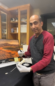
Dr. Sunil Manjila holding the multiport MR-compatible neuroendoscope prototype
Dr. Manjila with the prototype neuroendoscope

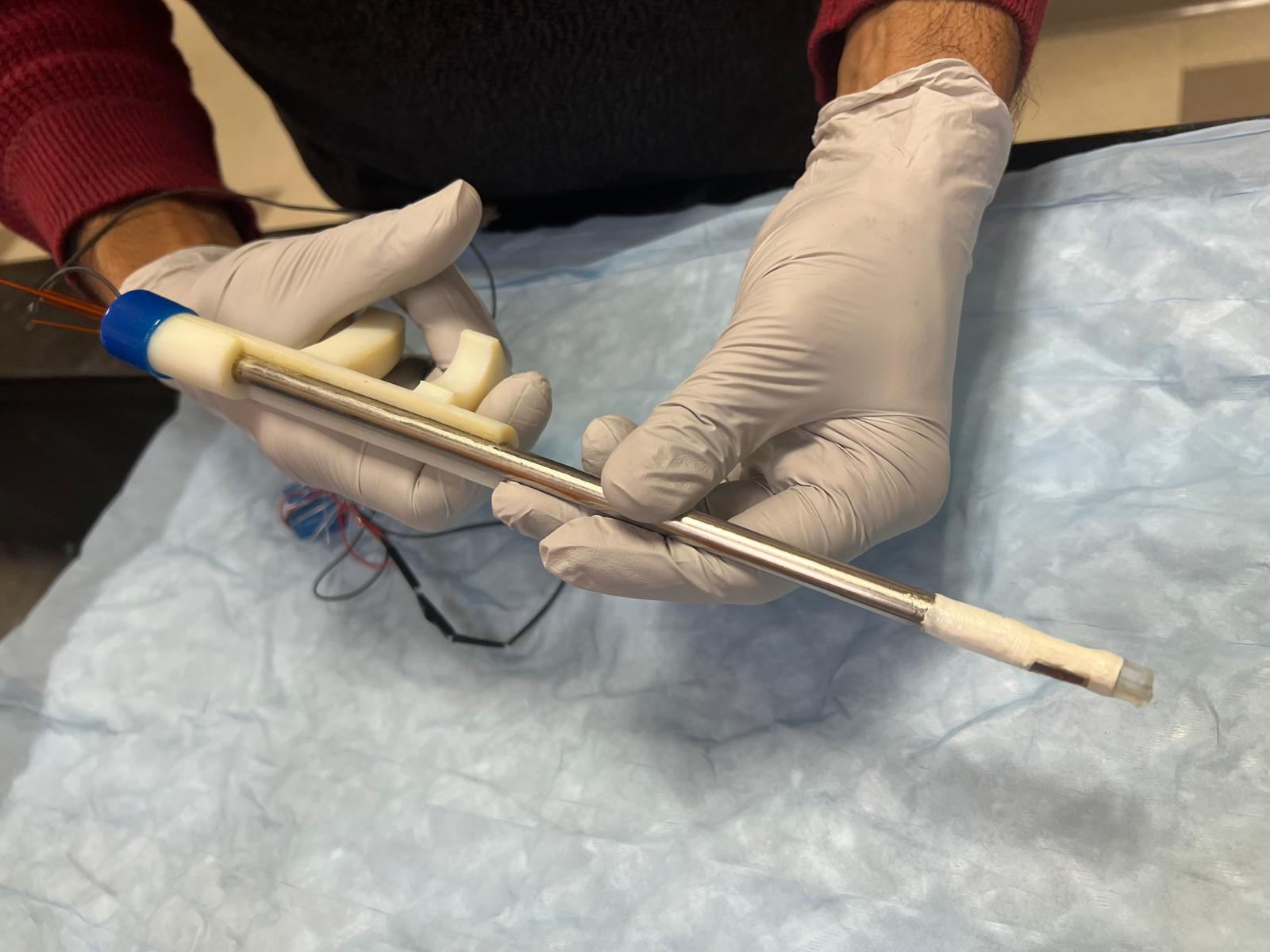
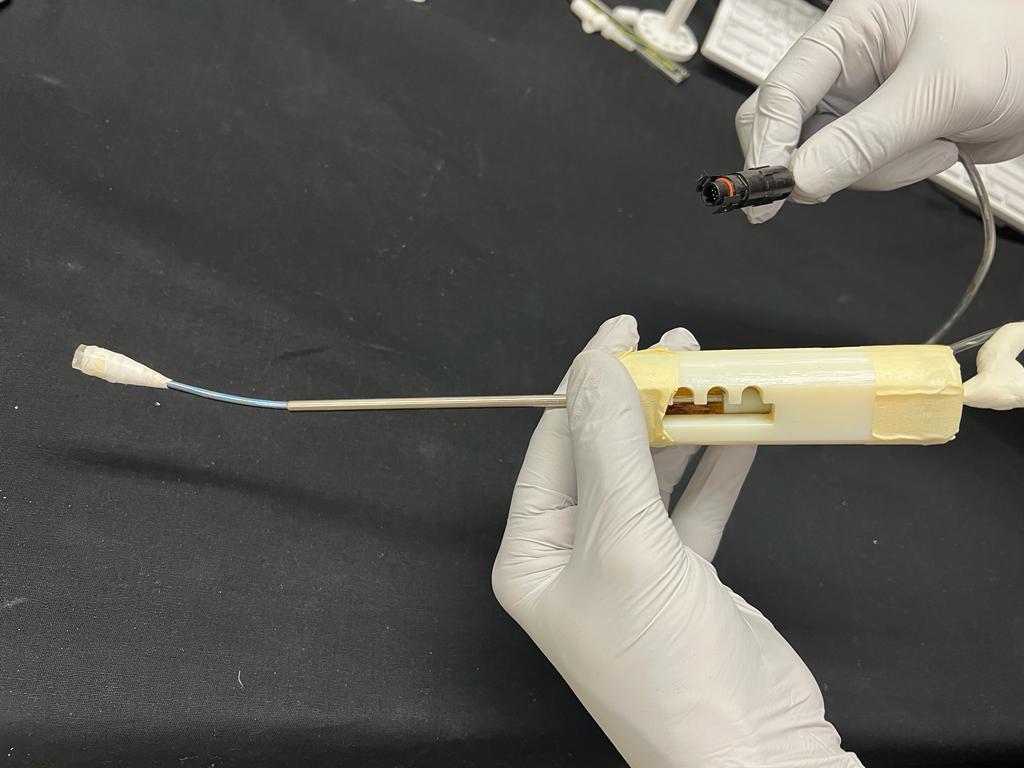
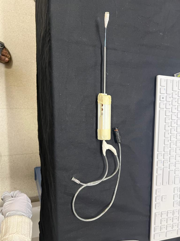
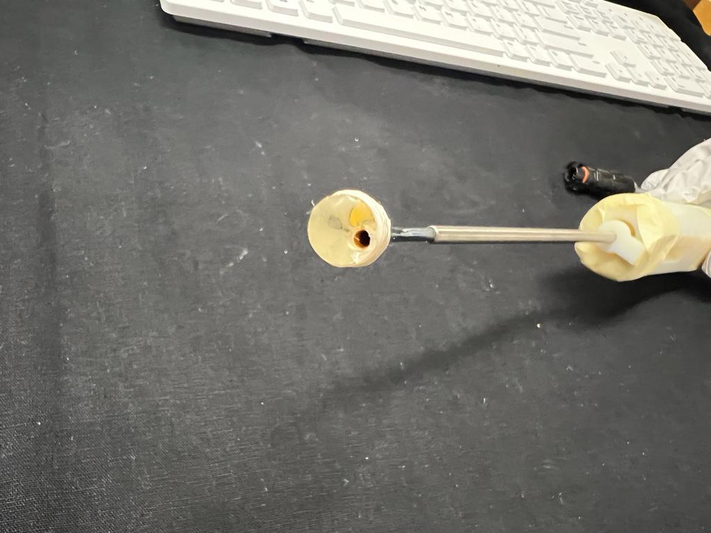
Conventional rigid endoscopes limit the surgeon to a single axis of vision at the instrument tip, requiring repeated rotation and risking tissue compression. Flexible scopes improve steerability but sacrifice channel capacity and control. Dr. Manjila's innovation bridges both — a hybrid rigid endoscope with two independent optical ports (tip and lateral), each with its own CMOS camera and LED illumination, plus a compliant silicone tip window that enables pre-deployment instrument visualisation and imaging in blood-filled fields.

Validated in porcine brain models, human cadaver studies, and inside a 3-Tesla MRI scanner, the prototype performed combined ventricular procedures with less than 5° of endoscope rotation. Its customisable port configurations, reduced brain distortion risk, and full MR compatibility open new frontiers — from laser-assisted tumour treatment to management of complex postinflammatory hydrocephalus — and form the basis of a granted US patent.

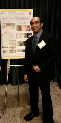
Dr. Sunil Manjila presenting the Soft-Touch Neuroendoscope poster
Presenting the Soft-Touch Neuroendoscope · Harvard Medical School
Innovation — image 1
Innovation — image 2
Innovation — image 3
Innovation — image 4

Principal Investigator: Pierre E. Dupont, PhD
https://robotics.tch.harvard.edu/

Selected Publications

Beyond the jugular bulb classification, Dr. Manjila's scholarship encompasses endoscopic cranial neurosurgery, skull base pathology, and the novel field of robotic cranial neurosurgery.

2023

Robotic Instruments Inside the MRI Bore: Key Concepts and Evolving Paradigms in Imaging-Enhanced Cranial Neurosurgery

World Neurosurgery · 2023 Aug;176:127-139

A comprehensive review of robotic cranial neurosurgery paradigms operating within MRI bore environments, examining imaging guidance, safety, and future directions.

DOI: 10.1016/j.wneu.2023.01.025 →
2016

A Multiport MR-Compatible Neuroendoscope: Spanning the Gap Between Rigid and Flexible Scopes

Neurosurgical Focus · 2016 Sep;41(3):E13

Introduced a novel multiport neuroendoscope design compatible with MRI environments, bridging the functional gap between rigid and flexible endoscopic systems — basis of a US patent.

DOI: 10.3171/2016.7.FOCUS16181 →
2019

Minimally Invasive Bilateral Anterior Cingulotomy via Open Minicraniotomy Using a Novel Multiport Cisternoscope: A Cadaveric Demonstration

Operative Neurosurgery · 2019 Feb;16(2):217-225

Demonstrated a minimally invasive surgical technique for bilateral anterior cingulotomy using a novel multiport cisternoscope, offering a cadaveric proof-of-concept for pain surgery approaches.

DOI: 10.1093/ons/opy083 →
Forthcoming

New Publication — Coming Soon

Details to be announced

A new paper by Dr. Sunil Manjila is currently in preparation. Details will be updated upon publication.

Contact

For academic inquiries, copyright or collaboration requests, or clinical correspondence regarding the Manjila-Semaan Classification.